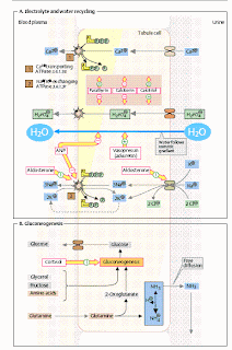
A. Electrolyte and water recycling _
Electrolytes and other plasma components with lowmolecular weights enter the primary
urine by ultrafiltration (right). Most of these substances are recovered by energy-dependent resorption (see p. 322).
The extent of the resorption determines the amount that ultimately reaches the final urine and is excreted. The illustration does not take into account the zoning of transport processes in the kidney (physiology textbooksmay be referred to for further details). Calcium and phosphate ions.
Calcium (Ca2+) and phosphate ions are almost completely resorbed from the primary urine by active transport (i.e., in an ATP-dependent fashion). The proportion of Ca2+ resorbed is over 99%, while for phosphate the figure is 80–90%. The extent to which these two electrolytes are resorbed is regulated by the three hormones parathyrin, calcitonin, and calcitriol. The peptide hormone parathyrin (PTH), which is produced by the parathyroid gland, stimulates Ca2+ resorption in the kidneys and at the same time inhibits the resorption of phosphate.
In conjunction with the effects of this hormone in the bones and intestines (see p. 344), this leads to an increase in the plasma level of Ca2+ and a reduction in the level of phosphate ions. Calcitonin, a peptide produced in the C cells
of the thyroid gland, inhibits the resorption of both calciumand phosphate ions. The result is an overall reduction in the plasma level of both ions.
Calcitonin is thus a parathyrin antagonist relative to Ca2+. The steroid hormone calcitriol, which is formed in the kidneys (see p. 304), stimulates the resorption of both calciumand phosphate ions and thus increases the plasma level of both ions. Sodium ions. Controlled resorption of Na+ from the primary urine is one of the most important functions of the kidney. Na+ resorption is highly effective, with more than 97% being resorbed. Several mechanisms are involved: some of the Na+ is taken up passively in the proximal tubule through the junctions between the cells (paracellularly). In addition,there is secondary active transport together with glucose and amino acids (see p. 322).
These two pathways are responsible for 60–70% of total Na+ resorption. In the ascending part of Henle’s loop, there is another transporter (shown at the bottom right),
which functions electroneutrally and takes up one Na+ ion and one K+ ion together with
two Cl– ions.
This symport is also dependent on the activity of Na+/K+ ATPase [2], which
pumps the Na+ resorbed from the primary urine back into the plasma in exchange for K+.
The steroid hormone aldosterone (see p. 55) increases Na+ reuptake, particularly in
the distal tubule, while atrial natriuretic peptide (ANP) originating from the cardiac atrium reduces it. Among other effects, aldosterone induces Na+/K+ ATPase and various
Na+ transporters on the luminal side of the cells. Water. Water resorption in the proximal
tubule is a passive process in which water follows the osmotically active particles, particularly the Na+ ions.
Fine regulation of water excretion (diuresis) takes place in the collecting ducts, where the peptide hormone vasopressin (antidiuretic hormone, ADH) operates. This promotes recovery of water by stimulating the transfer of aquaporins (see p. 220) into the plasma membrane of the tubule cells via V2 receptors. A lack of ADH leads to the disease picture of diabetes insipidus, in which up to 30 L of final urine is produced per day. B. Gluconeogenesis _ Apart from the liver, the kidneys are the only
organs capable of producing glucose by neosynthesis (gluconeogenesis; see p.154).
The main substrate for gluconeogenesis in the cells of the proximal tubule is glutamine.
In addition, other amino acids and also lactate, glycerol, and fructose can be used as
precursors. As in the liver, the key enzymes for gluconeogenesis are induced by cortisol
(see p. 374). Since the kidneys also have a high level of glucose consumption, they only
release very little glucose into the blood.


0 comments:
Post a Comment